
Tissue Culture of Fragaria (Strawberry)


The Fragaria: Strawberries
What comes to your mind when you hear “strawberry”? Definitely your favorite ice cream, the juicy fruit, or delicious cup-cake or thick shake!
Strawberries are one of the most consumed fruits all over the world. Their fragrance, taste, bright red color, juicy texture, and other nutritional properties make it appreciable among consumers. And, for this reason, it’s one of the most cultivated fruit crops worldwide.
The conventional techniques are not enough to meet the sky-high demands of market, processing, and export. Further, they are slow, laborious, and expensive with multiple challenges. This article talks about the in vitro technique of propagation of strawberries having a rapid and high multiplication rate.
Botany of the Strawberry
Strawberry, scientifically known as Fragaria, is a genus belonging to the Rosaceae family. The family harbors more than 20 species, hybrids, and cultivars. The plants are native to the northern hemisphere. Among all the varieties of strawberry, the garden strawberry (a cross of Fragaria x ananassa) is a widely grown species.
Some other major species of the genus Fragaria are F. moschata, F. vesca, F, chiloensis, and F. virginiana.
Strawberries are low-growing herbaceous plants. They have a fibrous root system and a crown from which basal leaves arise. The leaves of the plants are compound and hairy having edgy margins. The red or white-colored flowers of the plants are present in clusters. The fruits of strawberries are not true berries.
The fleshy and edible part of the fruit is referred to as receptacle (the stalk where flowers are attached) and the tiny embedded materials, called seeds, are achenes (true botanical fruits).
Strawberry propagation
Strawberry fruits are rich in vitamin C, B1, B2, protein, calcium, potassium, copper, and iron—the nutritious elements essential for the functioning of living organisms. Conventionally these plants are grown using stolon or runner segments of the plants. But, this is not an efficient technique for the production of plants at a commercial scale. Further, the number of propagules is limited when plants are propagated using stolon or runner and they are also susceptible to many diseases. It includes diseases caused by fungi, Strawberry mottle virus (SMV), and Strawberry mild yellow edge virus (SMYEV).
Here’s mentioned the procedure of strawberry micropropagation using shoot tip. The study is similar as mentioned in the book Tissue culture Techniques for Horticulture crops by Kenneth C. Torres with slight alteration at the callus maintenance stage.NOTE:The procedure mentioned here doesn’t entirely guarantee the successful propagation of plants. So, it’s highly recommended that culturists test the propagation with different concentrations of growth hormones and media components for the successful propagation of plants.

Procedure
Explant Preparation
- Collect shoot tips from the mother plant and peel them leaving only a couple of immature leaves covering the meristematic tissue.
- Surface sterilizes the explants by dipping them in 10% Clorox for 10 minutes. Then, rinse them twice using distilled water.
- Again peel the shoot tip leaving only the meristematic tissue.
- Resterilize the explant (meristematic tissue) in 5% Clorox for 5 minutes.
Shoot Initation
- Prepare shoot induction media with MS micronutrients containing 20 g/liter sucrose, 8 g/liter agar, and the following compounds:
| S. No. | Component | Concentration (mg/liter) |
| 1. | CaN03 | 1000 |
| 2. | KHzP04 | 250 |
| 3. | KN03 | 250 |
| 4. | MgS04 | 250 |
| 5. | Myo-inositol | 100 |
| 6. | Glycine | 2 |
| 7. | Nicotinic Acid | 0.5 |
| 8. | Pyridoxine | 0.5 |
| 9. | Thiamin | 1.0 |
| 10. | BA | 1.0 |
| 11. | IBA | 1.0 |
| 12. | GA3 | .10 |
- Dispense 10 ml media to each culture tube and sterilize them.
- After sterilization, cool the medium and inoculate each tube with one shoot tip.
- After inoculation, seal the tube using parafilm.
- Incubate the cultures at 25°C under a 16-hr photoperiod.
- To obtain increased growth transfer the explants to a fresh medium on a monthly period.
Related: Overview of Callus / Organ Tissue culture
- Prepare a fresh medium containing MS salts, 0.3% sucrose, and 8 g/liter agar. Sterilize the medium. Then add 0.5 mg/liter kinetin.
- Dispense 10 ml of media in each tube.
- Divide callus obtained from shoot initiation step into pea-sized tissues and culture each tissue in a single media-containing tube. And, incubate the plants at 25°C.
Rooting
- Prepare a rooting medium containing MS salts, 20 g/liter sucrose, 8 g/liter agar, and the following components:
| S. No. | Component | Concentration (mg/liter) |
| 1. | Myo-inositol | 100 |
| 2. | Glycine | 2.0 |
| 3. | Nicotinic Acid | 0.5 |
| 4. | Pyridoxine | 0.5 |
| 5. | Thiamin | 0.1 |
| 6. | IBA | 1.0 |
- Sterilize the medium.
- Dispense 10 ml of rooting medium to each tube.
- If shoots reached a height of 20 mm, excise them and transfer them to the rooting medium. In 1-2 months, profuse root growth will be observed.
- Incubate cultures at 25°C.
Hardening and Acclimation
- After roots are nicely formed, transfer the plants to a well-lighted area and a maintained temperature of 25°C for 2-5 weeks.
- Prepare a soil mixture containing 1 part jiffy mix, 1 part pine bark, and 1 part garden soil by volume.
- Transfer the mixture to a peat pot.
- Take out the plantlets from the media, wash the media attached to their roots, and transfer them to the peat pot.
- Water plantlets and spray with Benlate.
- Place potted plantlets in a covered plastic box to maintain high relative humidity. Gradually, increase the humidity by removing the plastics.
After a few weeks, you will observe healthy growing plants. And, if the protocol works for you, let us know at info@plantcelltechnology.com. We’ll be happy to feature your story on our social media platforms.
Happy Culturing!

Source: Giphy
Blog Categories
View by Level
Popular Blogs
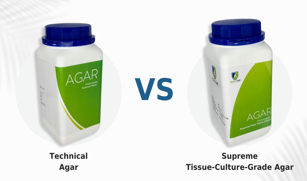
New Technical Agar Vs Supreme Agar
Introduction What’s the secret element that supports and holds plants in vitro? Not sure? It’s the solidifying agent. Solidifying agents...
Read More
Get the Protocol: How to Tissue Culture Nepenthes Using Nodes and Seeds
Introduction This plant is non-vegeterian... ...and we're not kidding! Nepenthes belongs to one of the most interesting families of carnivorous...
Read MoreFollow Us On Social


Follow Us On Social
Follow Us On Social
Join the conversation
Your email address will not be published. Required fields are marked