Tissue Culture Contamination and 7 Easy Steps of Prevention


Again, contamination!
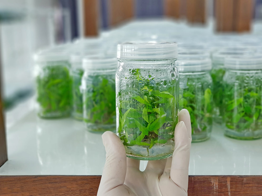
Tissue culture is a long and laborious process and it feels vexing when fungus or bacteria attack our lovely cultures.
Culturing cells in the labs requires a lot of effort, from the media preparation and cell transfer to the regular maintenance. But unfortunately, if the cells get contaminated and loss of cultures occurs, it results in a huge loss of time, effort, and money. The problem grows bigger if you don’t have any idea how your culture got contaminated and what kind of contamination it is!
It is devastating and you are left with no other choice but to repeat the experiments to save your research.

Types of Tissue Culture Contamination
Tissue culture contamination is categorized into two groups: Chemical contamination and Biological contamination.
Chemical Contamination
Chemical contamination occurs due to the presence of any non-living components that negatively affect the growth of your culture. The sources and types of chemical contamination are:
- Media: The cell culture media contains nutrients, reagents, and water which are the major sources of contamination.
- Serum: It is a nutrient boost for cell cultures. It contains proteins, hormone, and some growth factors which stimulate tissue growth. However, a variation in the concentration of the hormones and growth factors can be toxic to the cell cultures.
- Endotoxin: It is a byproduct of Gram-negative bacteria and found in water, sera, or other culture components. It can be quantified by Limulus Amebocyte Lysate assay (LAL).
- Plastic tubing and storage vessels: Chemical residue (detergents or disinfectants) in the uncleaned storage vessel leach into media when it is transferred to the vessels. Moreover, plasticizers can also affect the culture growth.
- Fluorescent light: It can photoactivate some media components, such as riboflavin and tryptophan, that release hydrogen peroxide and free radicals that are toxic to the cells.
Biological Contamination
Biological contamination is caused due to the presence of living organisms in the culture. Such organisms include easily identifiable bacteria, yeast, and molds or hard to detect viruses, protozoa, and mycoplasmas.
- Bacteria, yeast, and molds are ubiquitous in nature. So, they can easily sneak in, colonize, and flourish into the cell cultures. Antibiotics are used to avoid the culture of bacteria, however, some resistant strains can still grow into it.
- There are certain slow-growing very small or intracellular bacteria that are difficult to get noticed during routine checks of the cultures. They can cause serious harm to multiple cultures.
- Virus, protozoa, and mycoplasma being intracellular in nature are difficult to get identified. They not only destroy the whole cell culture but they also pose potential hazards to human health.
What are the best ways to avoid Contamination?
To prevent the contamination and save your culture and experiment you should follow the precautionary steps given below, “before and during” your tissue culture experiments:
- Wear gloves and a lab coat, and be organized: Have you ever thought you might be contaminating the cultured cells yourself? Yes, it’s true! A human body is loaded with bacteria. So, it’s best to wear gloves and your lab coat. It should be kept inside the lab and cleaned regularly. You can also list the thing you have to take to the facility room and this will help you to finish your experiments in one go.
- Use high-grade chemicals: Only use certified and high-grade media and serum in your experiments. Also, the water and media of the culture should be sterilized well.
- Sterilize lab equipment: Always autoclave the lab equipment, such as pipette tips, storage vessel, beaker, forceps, etc. Before that, make sure your equipment is autoclavable!
- Check the facilities: Regularly clean and sterilize the facility or room where cells are cultured. Check the working of the laminar flow and incubators as well. Wipe the laminar surface and other equipment with 70% alcohol before performing the experiment.
- Check the imported cells: If you are importing cells from a company or other labs, it’s best to check the cell by growing a culture for at least two weeks. This will help you to save your reagents and effort before you culture the cells on a large scale.
- Avoid cell exposure to non-sterilized surroundings: Always try to keep the cells in a sterilized environment. The transfer of cells from the incubator to hoods should be quick. If your cells have remained outside for a prolonged period, do keep them in a different hood. This will help you save your other cultures as well.
- Regularly check the cultured cells: You have done so much to protect your cells, and you don’t want to get your culture contaminated at the end! So, keep an eye on your cultures and watch them regularly.
Although, you will most likely be able to save your culture by following the above steps, it is equally difficult to be precise and consistent with the prescribed ‘best-practices’. By these methods, you can only avoid chemical contamination and to some extent biological contamination as well. But, as bacteria start developing resistance towards antibiotics, they no longer remain the best solution to avoid biological contamination in your culture.
Catering to all these challenges, and to ease your culture growing experience, we have specifically developed PPM™ (Plant Preservative Mixture), a one-stop solution to all your contamination problems. It will guard your cells from any type of contamination, whether its airborne, waterborne, or through human contact.
References:
- https://safety.fsu.edu/safety_manual/supporting_do...
- https://www.thermofisher.com/blog/cellculture/prev...
- https://www.sigmaaldrich.com/technical-documents/a...
- https://www.technologynetworks.com/cell-science/ho...
- https://study.com/academy/lesson/cell-culture-cont...
- https://www.sigmaaldrich.com/technical-documents/a...
Blog Categories
View by Level
Popular Blogs

6 Plant Tissue Culture Books to Keep Learning
Introduction Most of us are fans of books when it comes to learning a topic in detail and in a...
Read More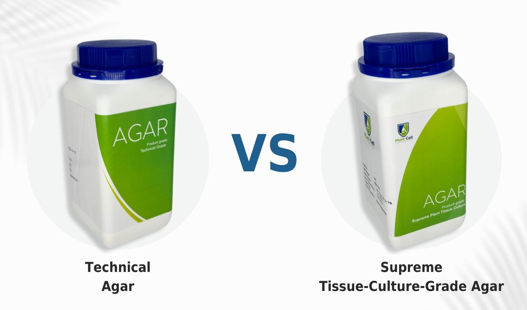
New Technical Agar Vs Supreme Agar
Introduction What’s the secret element that supports and holds plants in vitro? Not sure? It’s the solidifying agent. Solidifying agents...
Read MoreFollow Us On Social


Follow Us On Social
Follow Us On Social
Join the conversation
Your email address will not be published. Required fields are marked